Retinal Detachment: Guide to Symptoms and Treatments
Article by: Last updated: 14 July 2026

A sudden flash of light, an unexpected blur, or a dark shadow creeping across your vision can be highly alarming. These visual disruptions often signal a critical ophthalmic emergency known as retinal detachment. This condition occurs when the vital light-sensitive tissue at the back of your eye separates from its underlying support, putting your sight at immediate risk.
Understanding the crucial warning signs, underlying causes, risk factors, and advanced treatment options is essential to recognizing when to take immediate action to protect your lifelong vision.
Highlight
- Retinal detachment needs same-day ophthalmic evaluation, as delayed treatment, especially if the macula is affected, can reduce the chance of full vision recovery.
- Key symptoms include sudden flashes of light, a shower of dark floaters, or a curtain-like shadow over your visual field, all of which occur completely painlessly.
- Treatment relies heavily on advanced procedures like laser photocoagulation, cryopexy, or vitrectomy, tailored specifically to the severity and biological cause of the detachment.
- Individuals with high myopia, previous eye surgeries, chronic medical conditions, or a family history face an increased risk and require regular comprehensive dilated eye examinations.
Table of Content
Retinal detachment is a medical emergency where the retina, which is a thin layer of tissue at the back of the eye, pulls away from its normal position. This retinal detachment separation starves retinal cells of oxygen and essential nutrients. Left untreated, it can cause permanent vision loss. Key signs of retinal detachment include sudden flashes of light, new floaters, and a shadow over your visual field.
Yes, eye retinal detachment is a critical medical emergency requiring same-day care. Delaying treatment starves cells of oxygen, and if the detachment reaches the macula, central vision can be lost within days rather than being reversible. This raises a common question: how long before retinal detachment causes blindness? If you experience sudden flashes, floaters, or a curtain-like shadow, seek immediate emergency eye evaluation to save your sight.
While retinal detachment itself is entirely painless, critical warning signs often appear before it advances. Recognizing these visual disruptions and the specific symptoms of retinal detachment early is vital to saving your sight:
- The sudden appearance of tiny specks, hairs, or squiggly lines drifting across your field of vision, which shows what retinal detachment vision loss begins with.
- Noticing brief, bright sparks or bursts of light (photopsias) in one or both eyes, which are classic retinal detachment symptoms.
- Experiencing a sudden, unexplained loss of visual clarity.
- Gradual darkening or worsening of your side vision.
- A dark, shadow-like veil spreading over your visual field, which illustrates what does retinal detachment look like to an affected patient.
These retinal detachment causes primarily stem from aging changes or physical eye trauma. It is classified into three distinct biological types based on the underlying mechanism that forces the tissue away:
- Rhegmatogenous: The most common form, caused by a tear or hole in the retina that allows fluid to slip underneath and lift it.
- Tractional: Occurs when scar tissue grows on the retina’s surface, often due to diabetic retinopathy, and pulls it away.
- Exudative: An exudative retinal detachment is triggered by fluid accumulation beneath the retina without any tears, typically caused by inflammation, injury, or tumors.
While anyone can experience retinal detachment, specific factors significantly increase your vulnerability. Knowing if your profile falls into a high-risk category can save your eyesight through proactive screening, especially for those with high Myopia:
- Requiring strong glasses to see far away stretches and thins the retina.
- Prior eye procedures, particularly Cataract or glaucoma surgery, or other intraocular operations.
- History of a serious physical injury to the face or eye.
- Past retinal tears, detachment in your other eye, or genetics.
- Having thin, weak areas identified during an eye examination.
An ophthalmologist diagnoses retinal detachment through a dilated eye exam, using specialized drops to widen your pupil and inspect the retinal tissue. To view the internal structures with maximum precision, advanced imaging tools such as an ocular ultrasound or Optical Coherence Tomography (OCT) are utilized to detect microscopic tears or fluid accumulation.
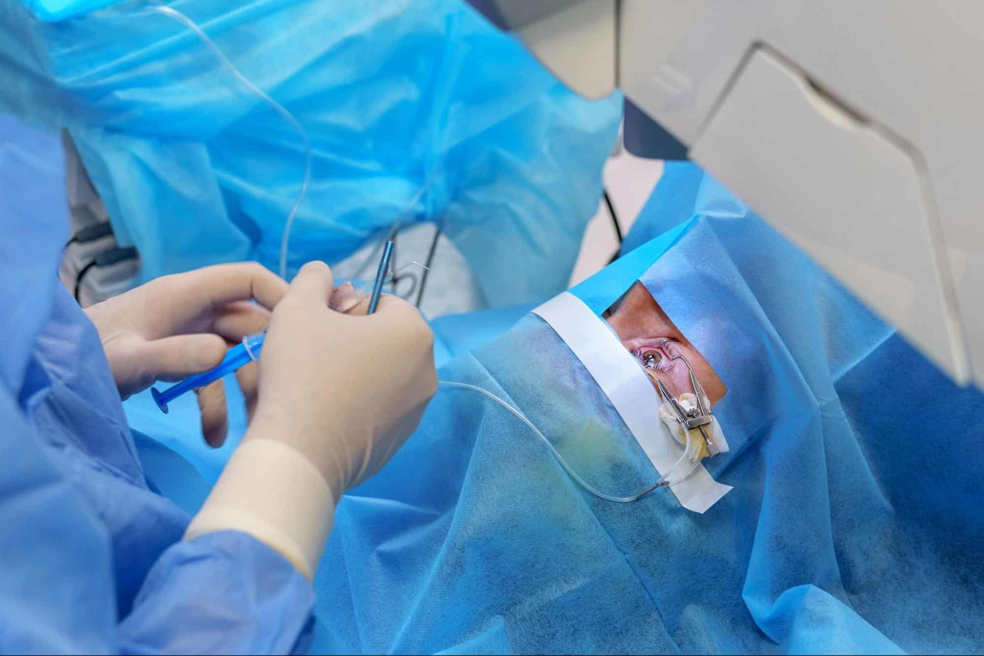
Retinal detachment cannot heal on its own and requires a specialized retinal detachment treatment. Your ophthalmologist will select the most effective retinal detachment surgery approach based on the size, location, and specific type of your retinal detachment:
Laser Photocoagulation
This highly precise procedure treats early-stage retinal tears before complete detachment occurs. An ophthalmologist directs a specialized medical laser through your dilated pupil, creating tiny, controlled burns around the torn tissue. As these burns heal, they form secure microscopic scars that effectively weld and bond the retina back down to its underlying support layer, preventing further fluid leakage.
Cryopexy
Cryopexy utilizes extreme cold to seal retinal tears and prevent advanced detachment. The surgeon applies a specialized, freezing probe to the outside wall of the eye directly over the site of the tear. This intense cold creates a localized scar around the damaged area, acting as a secure anchor that tightly holds the retinal tissue in its proper place.
Pneumatic Retinopexy
Pneumatic Retinopexy is a minimally invasive procedure involving injecting a gas bubble directly into the vitreous cavity of your eye. The bubble expands and gently presses the detached retina back against the eye wall. To ensure the bubble remains in the correct healing position, you must maintain a strict, specific head posture for several days as directed by your doctor.
Scleral Buckle
In the Scleral Buckle method, a tiny, flexible band made of silicone or soft plastic is permanently sewn onto the outside of the white part of your eye (the sclera). This band gently indents the eye wall inward, relieving the internal pulling forces and allowing the detached retina to reattach and heal securely against the underlying tissue.
Because the buckle applies gentle external pressure on the eye, it can temporarily change your cornea's curvature and induce astigmatism, usually resolving within a few months as the eye adjusts.
Learn more about astigmatism and how it's corrected: Astigmatism: Symptoms, Causes and Treatment Options
Vitrectomy
This comprehensive surgery involves removing the gel-like vitreous humor that is pulling on the retina. The surgeon then fills the eye with an air, gas, or silicone oil bubble to hold the retina in place. If gas is used, patients must strictly avoid flying, high altitudes, or scuba diving, as pressure changes can cause dangerous eye pressure spikes.
Understanding the potential complications and recovery requirements helps you make an informed decision alongside your ophthalmologist:
- Risks include eye infections like keratitis, internal bleeding, accelerated cataract development, or elevated eye pressure (glaucoma).
- The retina may fail to reattach properly, or redetach, requiring a second surgery. Studies consistently show single-surgery success rates around 90%, meaning roughly 1 in 10 patients need an additional procedure.
- Permanent blurry vision with retinal detachment reduction can persist even after anatomically successful reattachment.
- Recovery demands wearing an eye patch, resting for weeks, and knowing the things to avoid with retinal detachment recovery, such as maintaining strict head positioning if a gas bubble was used.
Your visual recovery depends heavily on whether the macula (the center of vision) was detached (macula-off) or intact (macula-on), and how quickly you received treatment. Understanding the patient's condition before and after retinal detachment eye surgery is crucial; macula-on cases yield much faster, better visual outcomes, whereas macula-off recovery can take months.
Essential aftercare rules for managing life after retinal detachment surgery include:
- Maintaining a specific head posture for 1–2 weeks to keep the gas bubble in place.
- Avoiding air travel, high altitudes, and scuba diving until the gas bubble completely dissolves to prevent dangerous eye pressure spikes.
- Attending all post-op check-ups to monitor healing and protect your sight.
Incorporating these protective measures into your lifestyle helps preserve long-term retinal health, especially if you have had a prior Phaco procedure or Cataract Surgery:
- Wear Protective Eyewear: Guard against physical trauma during high-risk sports or hazardous work.
- Annual Eye Exams: Schedule comprehensive dilated exams yearly to detect early microscopic tears.
- Manage Chronic Conditions: Keep blood sugar and blood pressure stable to prevent vascular damage.
- Maintain Eye Hygiene: Prevent severe infections or inflammation that could compromise retinal stability.
- Act Fast: Seek immediate ophthalmological care at the very first sign of visual changes.
Retinal detachment is a severe, painless medical emergency that demands rapid diagnosis and specialized surgical intervention to prevent permanent blindness. While biological factors like aging or high myopia increase your vulnerability, recognizing early warning signs like sudden floaters and flashes allows for timely treatment. Safeguarding your vision requires a combination of routine eye check-ups, proper protection, and immediate clinical action.
Samitivej Chinatown Hospital provides world-class ophthalmic care with a dedicated team of eye specialists and state-of-the-art diagnostic imaging. Whether you require immediate laser photocoagulation or advanced vitreoretinal surgery, our international-standard facility offers prompt, comprehensive treatments. Trust us as your dedicated partner in preserving your vision and ensuring a safe, seamless recovery journey.
What are the early warning signs of a detached retina?
The primary signs are a sudden shower of dark floaters, flashes of light, and a dark, curtain-like shadow spreading across your field of vision. Seek immediate emergency care if these occur.
Can retinal detachment heal?
No, a detached retina cannot heal on its own. It is a critical medical emergency that requires prompt surgical intervention to reattach the tissue and prevent permanent, irreversible blindness.
Can glasses correct vision after retinal detachment surgery?
Yes, glasses can often help optimize your remaining vision once the eye heals. However, final clarity depends heavily on whether the macula was damaged before surgery took place.