Anterior Cruciate Ligament: Anatomy, Symptoms & Treatment
Article by: seoteam seoteam Last updated: 9 September 2026
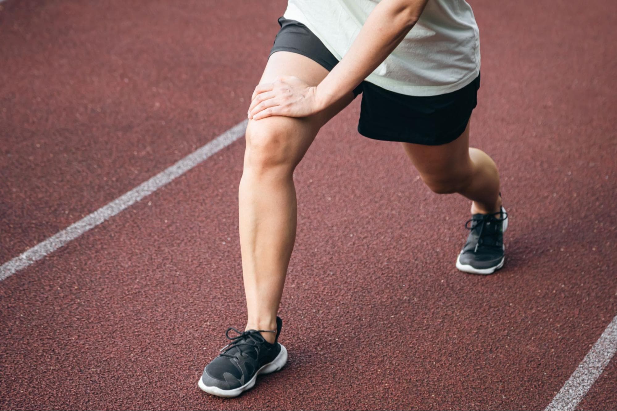
The anterior cruciate ligament (ACL) is one of the most vital stabilizing structures in the human knee joint, working continuously to control leg rotation and prevent the shinbone from slipping forward. Whether you are an athlete performing explosive pivots or simply staying active, maintaining the integrity of this ligament is key to overall mobility.
Understanding how the ACL functions alongside the common causes of tears, diagnostic methods, and modern recovery options, empowers you to make informed decisions about your joint health and long-term athletic performance.
Highlight
- The anterior cruciate ligament prevents the shinbone from sliding excessively forward beneath the thighbone and controls rotational stability during dynamic movements.
- Most ACL tears occur without direct physical impact, usually resulting from sudden stops, awkward landings, or sharp direction changes.
- Care ranges from conservative physical therapy for low-demand, partial tears to arthroscopic reconstruction surgery for active individuals.
- Returning to sports safely typically takes 6 to 12 months of progressive physical therapy and objective functional testing.
- Lower-body strength conditioning, proper landing mechanics, and neuromuscular training significantly reduce the risk of ligament injury.
Table of Content
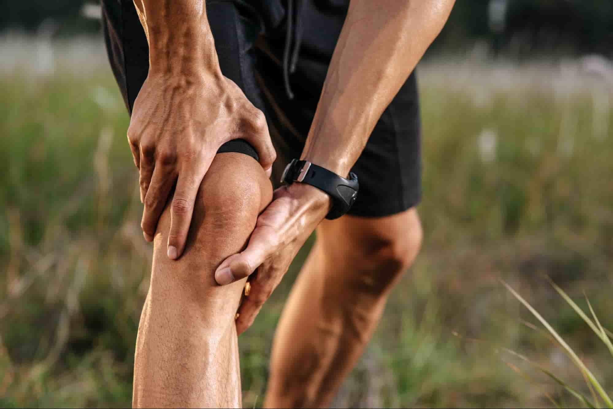
The anterior cruciate ligament (ACL) is a major stabilizing ligament located in the center of the knee joint. If you wonder what is the ACL or what is an ACL, it plays a critical role in controlling leg movements, preventing the shinbone from sliding forward beneath the thighbone, and providing dynamic rotational stability. When considering what is ACL or what does ACL stand for, the ACL full form and ACL acronym mean anterior cruciate ligament (known in Spanish as ligamento cruzado anterior).
Because of its key ACL action, it locks the knee in place during sharp cuts, pivoting, and twisting, making this ACL ligament especially crucial for athletes participating in high-impact sports like football. Understanding the anterior cruciate ligament meaning is essential for any athlete experiencing severe knee pain.
Where the ACL Is Located in the Knee
The anterior cruciate ligament (ACL) is situated deep within the center of the knee joint. For those asking where is your ACL, where is the ACL, where is ACL, or where is your ACL located, its ACL location runs diagonally from the back of the thighbone (femur) to the front of the shinbone (tibia). It forms an "X" shape alongside the posterior cruciate ligament (PCL) to connect and stabilize both long bones inside the ACL knee. (Note: unlike leg structures, there is no such thing as an ankle ACL, as this ligament belongs strictly to the knee).
Why the ACL Matters for Knee Stability
The ACL serves as the knee's primary internal stabilizer. By controlling the forward movement and rotational pivot of the tibia (shinbone) relative to the femur (thighbone), it keeps the joint firmly aligned throughout the ACL leg. Without a functional ACL, the knee becomes unstable, giving out during basic turning, stopping, or jumping motions.
An ACL injury typically occurs when severe, sudden rotational or impact forces stress the knee joint beyond its structural capacity. While commonly associated with high-impact sports, these injuries can happen during a variety of sudden physical movements
- Sudden Stops and Direction Changes: Rapidly decelerating and pivoting while running, cutting, or turning sharply.
- Awkward Landings: Landing improperly or with an extended knee after jumping.
- Direct Blunt Impact: Receiving a hard blow to the side of the knee joint, such as during a football tackle.
- Joint Overextension: Hyperextending the knee joint past its natural range of motion.
- Combined Structural Trauma: Suffering concomitant damage to nearby stabilizing structures, frequently occurring alongside medial collateral ligament (MCL) or meniscus tears.
While an ACL trauma can strike anyone during physical activity, certain demographics, physical traits, and athletic participation patterns significantly elevate an individual's overall vulnerability.
- High-Impact Sports Athletes: Competitors in sports involving rapid deceleration, pivoting, and jumping such as soccer, basketball, football, and downhill skiing.
- Female Athletes: Women experience anterior cruciate ligament tears up to four to six times more frequently than men due to differences in neuromuscular control, pelvic alignment, joint laxity, and hormonal influences.
- Individuals with Prior Knee Injuries: A history of knee trauma or prior ligament tears substantially increases the likelihood of secondary injury or re-tearing the ACL graft.
- People with Inadequate Physical Conditioning: Weak quadriceps and hamstrings, along with poor muscular balance or improper landing mechanics, put excessive stress directly onto the knee ligaments.
- Athletes Playing on Specific Surfaces: Exercising on high-friction surfaces, such as artificial turf or hard indoor courts, increases foot fixation during sudden twists, transferring severe torque to the knee joint.
An ACL tear typically presents immediately following a traumatic knee event, often leaving the ACL torn. Recognizing these acute physical indicators of an ACL tear knee is crucial for seeking timely medical evaluation
- Audible "Pop": Hearing or feeling a distinct popping sensation inside the knee at the moment of impact or direction change.
- Rapid Swelling: Marked joint swelling develops rapidly, often within 2 to 6 hours after the injury occurs.
- Severe Pain and Weight-Bearing Difficulty: Intense discomfort that makes placing weight or walking on the affected leg extremely painful.
- Joint Instability: A distinct feeling that the knee is weak, bucking, or "giving way" during basic movement.
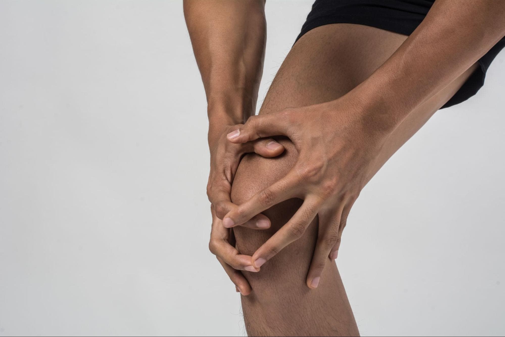
Accurately diagnosing an anterior cruciate ligament injury combines a careful medical history review with targeted clinical assessments and advanced diagnostic imaging to determine structural damage and evaluate overall joint stability.
Physical Examination Tests
During the initial consultation, your doctor performs a physical exam by inspecting the knee for visual swelling, bruising, and range of motion. If an ACL sprain or tear is suspected, specialized hands-on stability tests, such as the Lachman, Anterior Drawer, and Pivot Shift tests, are conducted to check for abnormal joint looseness by manually moving the shinbone forward relative to the thighbone.
Imaging: MRI and X-ray
Imaging confirms the physical findings. While X-rays cannot show soft tissue, doctors use them first to rule out associated bone fractures or cartilage detachment.
Magnetic Resonance Imaging (MRI) is the gold standard for diagnosis; it uses magnetic fields to produce detailed 3D pictures of internal knee structures, confirming whether a partial ACL tear or a complete tear has occurred while assessing nearby menisci, ligaments, and cartilage.
For patients with an isolated anterior cruciate ligament injury without accompanying tissue tears or structural knee damage, non-surgical management can be an effective approach—particularly for older individuals or low-demand patients whose daily activities do not place heavy stress on the knee. Treatment typically focuses on pain relief, anti-inflammatory medications, and structured physical therapy.
While physical therapy cannot naturally repair or restore a torn ACL, it strengthens the surrounding leg muscles to compensate for the lost ligament, providing functional knee stability that allows patients to manage daily tasks without surgery.
Surgical intervention is generally recommended when an anterior cruciate ligament tear occurs alongside other knee injuries, such as meniscal tears or damage to multiple collateral ligaments. It is also advised for younger individuals, athletes, or active professionals like military personnel and police officers whose lifestyles demand robust joint stability, as well as patients at high risk for premature knee osteoarthritis without intervention. Note that ACL reconstruction replaces a single ligament and is completely different from total knee arthroplasty, which replaces the entire joint surface.
Modern ACL reconstruction is performed as a minimally invasive arthroscopic procedure. Surgeons graft healthy tissue, typically harvested from the patellar tendon or nearby hamstring tendons, to create a functional replacement for the torn ligament. Arthroscopy ensures smaller incisions and faster recovery times, allowing patients to regain full knee function when paired with structured post-operative physical therapy.
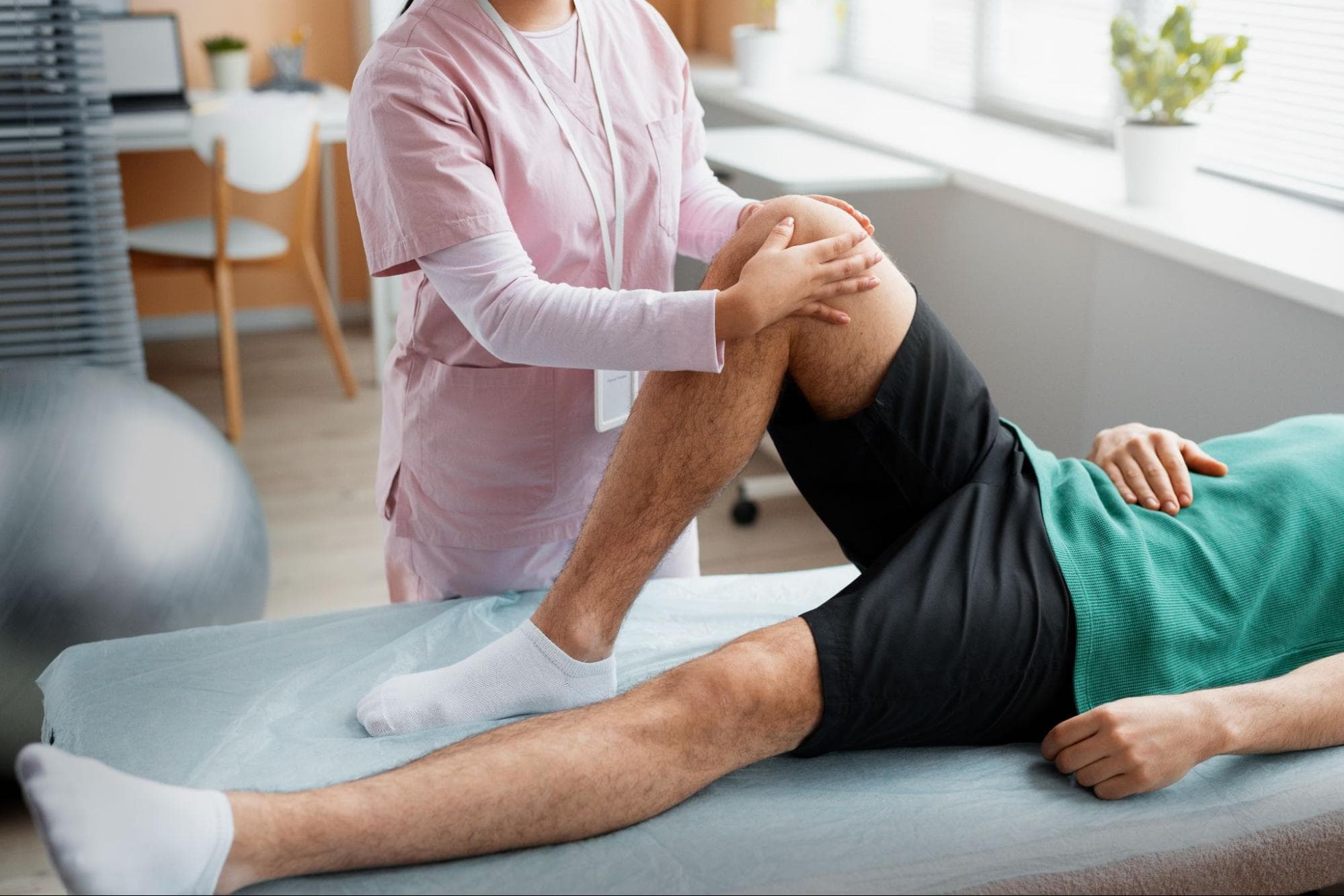
Recovering from an anterior cruciate ligament injury is a structured, multi-phase process requiring dedicated rehabilitation. Whether managed non-surgically or through operative reconstruction, a guided physical therapy protocol is essential to restore joint function, rebuild muscle strength, and ensure safe mobility.
Typical Recovery Timeline
Recovery from anterior cruciate ligament reconstruction generally spans 6 to 12 months, though consider "up to 24 months" for a safer margin, or soften to "at least 18 months" rather than presenting 18 as the endpoint. Early phases (weeks 1–6) focus on pain management, swelling reduction, and regaining full passive knee extension.
Mid-stage rehabilitation (months 2–4) emphasizes progressive quadriceps and hamstring strengthening, dynamic balance exercises, and restoring natural gait mechanics.
Physical Therapy and Return-to-Sport Testing
Physical therapy progresses from basic range-of-motion drills to high-level neuromuscular and plyometric training. Before returning to competitive play, athletes must pass comprehensive functional assessments including single-leg hop tests, agility drills, and strength asymmetry evaluations.
Clearing these objective benchmarks rather than relying solely on time post-surgery significantly lowers the risk of secondary graft re-rupture. Patients often share their recovery experiences and support tips on online communities like r/acl.
Protecting the anterior cruciate ligament is vital for maintaining overall knee stability and preventing secondary joint damage. Implementing targeted preventive strategies can significantly reduce your injury risk
- Targeted Lower-Body Conditioning: Build strength in the knee and leg muscles to reinforce joint stability and absorb impact forces.
- Professional Sports Coaching: Work with sports scientists or qualified trainers to learn correct technique and body positioning during movement.
- Proper Landing and Movement Mechanics: Practice controlled jumping, landing, and cutting techniques to avoid placing excess stress on the ligament during high-impact sports.
Navigating an anterior cruciate ligament injury requires a thorough understanding of joint stability, accurate diagnostic testing, and a personalized treatment strategy. Whether electing conservative rehabilitation or modern arthroscopic reconstruction, committing to structured physical therapy remains key to restoring mobility, preventing secondary joint wear, and safely returning to active living.
Don't let knee pain or instability hold you back from living fully. The experienced sports medicine specialists and orthopedic surgeons at Samitivej Chinatown Hospital offer comprehensive diagnostic evaluations, tailored treatment plans, and advanced rehabilitation services. Schedule a consultation with our orthopedic team today to get back on track safely.
How long does it take to heal from an anterior cruciate ligament tear?
Full recovery typically takes 6 to 12 months, depending on whether you undergo surgery or conservative rehabilitation. While basic walking may resume within weeks, full return to high-impact sports requires passing objective physical strength and stability benchmarks.
Is an ACL tear the same as an ACL sprain?
Yes, medically speaking. An ACL sprain is a broad diagnostic category for ligament stretching or tearing, graded from 1 to 3. A Grade 3 sprain represents a complete tear of the ligament fibers, requiring similar medical evaluation.
Can children and teenagers get ACL injuries?
Yes, growing athletes frequently suffer ACL tears during high-impact sports. Pediatric cases require specialized surgical techniques—such as physeal-sparing reconstruction—to repair the ligament without damaging active growth plates or affecting future bone development in young patients.
Is a knee brace necessary after an ACL injury?
A brace is often used initially to stabilize the joint, restrict harmful movements, and protect healing tissues post-surgery. However, long-term recovery relies primarily on targeted physical therapy to rebuild surrounding muscular strength rather than permanent brace dependence.
Can you re-injure your ACL after treatment?
Yes, re-injury can occur in either the surgical graft or the opposite knee. Returning to sports prematurely, inadequate muscle strength, poor landing mechanics, and skipping post-operative physical therapy significantly increase the risk of secondary ACL tears.